Importance of Splenectomy in SCD Patients
Reticuloendothelial System :
– Reticuloendothelial system also called as tissue macrophage system is the system of primitive phagocytic cells.
– Reticuloendothelial system is found in following structures :
1) Endothelial lining of vascular & lymph nodes, channels.
2) Connective issue & some organs like spleen, liver, lungs, lymph
nodes, bone marrow.
Reticular cells in these tissues form the tissue macrophage system.
Classification of Reticuloendothelial cells :
1) Fixed Reticuloendothelial cells or tissue macrophages.
2) Wandering Reticuloendothelial cells.
1) Fixed Reticuloendothelial cells or tissue macrophages.
– These cells are also called as fixed histiocytes for they are located in tissues.
– Tissue macrophages are present in following areas :
(i) Connective Tissues – Reticuloendothelial cells in connective tissues & in serous membranes like pleura, omentum & mesentaries are called fixed macrophages of connective tissue.
(ii) Endothelium of Blood Sinusoid :- In bone marrow, liver, spleen, lymph nodes, adrenal glands and pituitary glands also contain fixed cells. Kupffer cells in liver belong to this category.
(iii) Reticulum :- Reticulum of spleen, lymph nodes, bone marrow contain fixed reticuloendothelial cells.
(iv) CNS :- Meningocytes & meninges & microglia form tissue macrophages of brain.
(v) Lungs :- Tissue macrophages are present in alveoli of lungs.
2) Wandering Reticuloendothelial cells & tissue macrophages :
– Also called as Free histiocytes.
– These are of two types.
(i) Free Histiocytes of blood :
1) Neutrophils
2) Monocytes which become macrophages & migrate to site of
injury or infection.
(ii) Free Histiocytes of Solid Tissue :
During emergency, fixed histiocytes from connective tissue & other organs become wandering cells and enter the circulation.
– “Reticuloendothelial system” is an older term for the mononuclear phagocyte system, but it is used less commonly now, as it is understood that most endothelial cells are not macrophages.
– Spleen is the largest unit of mononuclear phagocyte system.
Monocyte is formed in bone, marrow & transported by blood it migrates into tissues where it transforms into histiocyte or a macrophage.
– Macrophages are diffusely scattered in connective tissue, liver (kuppffer cells), spleen & lymph nodes, lungs & CNS.
Functions of Reticuloendothelial System :
– Reticuloendothelial System plays an important role in defense mechanism of body.
– Most of the functions of RE system are carried out by the tissue macrophages.
– Spleen is major component of Reticuloendothelial system, producing lymphocytes in newborn and containing phagocytes, which remove worm-out RBC’s & other foreign bodies from bloodstream.
Functions of Tissue Macrophages are :-
1) phagocytic Function :- Macrophages are large phagocytic cells, which play an important role in defense mechanism of body, by phagocytosis.
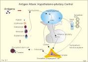
When any foreign body invades, macrophages ingest them by phagocytosis and liberate the antigenic products of organism. Antigens activate the helper T-lymphocytes & B-lymphocytes.
Lysosomes of macrophages contain proteolytic enzymes and Lipases, which digest bacteria & other foreign bodies.
2) Secretion of Bactericidal Agents :-
Tissue macrophages secrete many bacteriocidal agents which kill the bacteria. The important bactericidal agents of macrophages are oxidants. An oxidant is a substance that oxidizes another substance.
Oxidants secreted by macrophages are :-
Superoxide (O-2)
Hydrogen peroxide (H2O2)
Hydroxyl ions (OH-)
These oxidants are most potent bactericidal agents. So, even the bacteria which cannot be digested by lysasomal enzymes are degraded by these oxidants.
3) Secretion of Interleukins :-
Tissue macrophages secrete the following interleukins which help in immunity :
Interleukin – 1 – Accelerates the matunation and proliferation of specific B-lymhocytes & T-lymphocytes.
Interleukin – 6 – causes growth of B-lymphocytes & production of antibodies.
Interleukin – 12 – Influences the T-helper cells.
4) Secretion of Tumour Necrosis Factor :-
Two types of Tumour Necrosis Factor are secreted by Tissue macrophages.
TNF – : Causes necrosis of Tumour and activates immune responses in body.
TNF – : Stimulates immune system and vascular response, in addition to causing necrosis of tumour.
5) Secretion of Transforming Growth Factor :-
Tissue macrophages secrete Transforming agents i.e. growth factors which play on important role in preventing rejection of transplanted tissues or organs by immunosuppression.
6) Secretion of Platelet – derived Growth Factor :-
Tissue macrophages secrete the platelet-derived growth factor or PGDF which accelerates repair of damaged blood vessels and wound healing too.
7) Removal of Carbon Particles and Silicon :-
Macrophages ingest the substances like carbon dust particles (CO, CO2) and silicon, which enters the body.
8) Destruction of senile Aged RBC :-
Reticuloendothelial cells, particularly those in spleen destroy the senile RBC’s and release the haemoglobin.
9) Destruction of Haemoglobin :-
Haemoglobin released through broken senile RBC’s is degraded by the reticuloendothelial cells.
10) Storage functions :-
A large amount of lipids, cholesterol & iron are stored in Reticulo-Endothelial cells.
11) Formation of bile pigments :-
RE cells manufacture bilirubin from haemoglobin.
Spleen as a part of Reticulo Endothelial System :-
‘Kupffer cells’ are found in liver. Some sort of cells in brain are called microglia. While in spleen they are called as ‘reticular cells’.
Spleen’s Special Role :-
– The spleen usually has a particularly vital function in RES. This organ is specially designed to filter blood, making sure that deformed and old cells are removed from circulation & broken down while maintaining the haemoglobin count of Red blood cells.
– Spleen and its cell play an important role in immune function & disease and infection prevention.
– When the cells in the RES grow confused or tired they are more likely to make mistakes which can lead to more infections, waste build up in blood, and a number of potentially serious complications.
– Spleen is a central player in reticuloendothelial system.
– Specialized macrophages residing in spleen, breakdown old RBC’s and metabolize the haemoglobin, thereby freeing the iron compound haeme for production of new RBC’s.
Internal Structure of Spleen :-
Spleen is the largest lymphoid organ in the body & it is highly vascular. The spleen combines the innate & adaptive immune system. The structure of spleen enables it to remove older erythorocytes from the circulation & leads to the efficient removal of blood-borne microorganisms.
In essence the spleen is organized as a ‘free’ of branching arterial vessels, in which the smaller arterioles end in a Venous Sinusoidal System. The organ is surrounded by fibrous capsule of connective tissue, stemming from which are Trabecule that support the larger vasculature.
In humans part of bloodstream ends in the perifollicular zone with its locations in the circulatory system & with the unusual structure of its lymphoid compartment, the spleen is unique lymphoid organ. The combination of highly adapted macrophages & specific anatomical features of the marginal zone in particular underlies the fact that the spleen is crucial site of early exposure to encapsulated bacteria.
Schema of the spleen The afferent splenic artery branches into central arterioles which are sheathed by white pulp areas, these white pulp areas consist of T-cells zone. (also known as periarteriolar lymphoid sheath), arterioles & B-cells follicles. The arterioles end in cords in the red-pulp from where the blood runs into the venous sinuses, which collect into the efferent splenic vein. The larger arteries & veins run together in connective tissue trabeculae, which are continuous with the capsule that surrounds the spleen.
Formation of Blood Cells :-
Spleen plays an important role in the hemopoietic function in embryo. During the hepatic stage, spleen produces blood cells along with liver. In myloid stage, it produces the blood cells along with liver & bone marrow.
Destruction of Blood Cells :-
Older RBCs, lymphocytes & thrombocytes are destroyed in the spleen, when RBCs become old, the cell membrane becomes more fragile.
Diameter of most of the capillaries is less or equal to that of RBC. The fragile old cells are destroyed while trying to squeeze through the capillaries because these cells cannot withstand the stress of squeezing.
Destruction occurs mostly in capillaries of spleen because the splenic capillaries have a thin lumen, so the spleen is known as ‘graveyard of RBCs’.
Splenic pulp :-
The parenchymal tissue which is enclosed within capsule is the splenic pulp. It has 2 distinct types (1) white pulp (2) red pulp.
1) White pulp –
In a freshly sectioned spleen the white pulp are seen scattered all throughout the red pulp as grey patches. These grey patches at early periods were described as Malpeighian bodies.
The white pulp is accumulation of lymphatic tissue surrounding a major arterial vessels of the spleen. This lymphatic tissue is comprised of lymphocytes, plasma cells, macrophages or other free cells lying in the meshwork of reticular fibers.
In the white pulp there are two distinct components are seen.
1) As the arteries leave the trabeculae & enter splenic parenchyma, the vessels lose its adventitia & are replaced by reticular tissue followed by invasion with lymphocyte. This constitutes the periarterial lymphatic sheath of the white pulp.
2) At various points along the courses of vessels where the infiltration of lymphocytes is greater it forms ovoid nodules which are called as splenic nodules of white pulp.
2) Red Pulp :-
It is modified lymphatic tissue & is mostly infiltrated with cells of circulating blood. It consist of two components.
1) Splenic sinuses or sinusoids –
Splenic sinuses are long vascular channels having 35 to 40 in diameter. They may have an irregular course & may vary in diameter. They extend throughout the red pulp.
2) Splenic cords :-
Splenic cords appear as continuous partitions in between the sinuses. These cellular cords ultimately form a spongy network of modified lymphatic tissue that gradually merges into the white pulp.
In mammalian embryos the red pulp contains myelocytes erythroblasts & also megakaryocytes. These types of cells are not present in adult spleen except in certain pathological conditions.
Marginal Zone :-
It is the functional organ in between the white pulp & red pulp & consists of meshwork of branched reticular cells in association with extra cellular reticulum, into which many arterial vessels open.
Sinusoid of the spleen :-
A sinusoid is a small blood vessel that is a type of capillary similar to a fenestrated endothelium.
Sinusoids are actually classified as a type of open pore capillary as a opposed to continuous & fenestrated types, fenestrated capillaries have diaphragms that covers the pores whereas open pore capillaries lack a diaphragm & just have an open pore.
The open pores of endothelial cells greatly increases their permeability. In addition permeability is increased by large inter-cellular clefts & fewer fight junctions. The level of permeability is such as to allow small & medium sized proteins such as albumin to readily enter & leave the blood stream.
Sinusoids are found in the liver, lymphoid tissue, endocrine organs & hematopoietic organs such as the bone marrow & the spleen.
Sinusoids found within terminal villi of the placenta are not comparable to these because they possesses a continuous endothelium & complete basal lamina. This word was first used in 1893.
Phagocytic function of the spleen :-
Phagocytosis – The spleen is an important component of the reticuloendothedial system. The splenic phagocytes include-
1) The reticular cells & free macrophages of the red pulp.
2) Modified reticular cells of the ellipsoids.
3) free macrophages & endothelial cells of the venous sinusoids
4) Surface reticular cells of the lymphatic follicle.
The phagocytes present in the organ remove cell debris & odd & effete RBCs, other blood cells & microorganisms & thus filter the blood.
5) Phagocytosis of circulating antigens initiates humoral & cellular immune responses.
6) As a part of mononuclear phagocyte system, it metabolizes hemoglobin removed from senesent erythrocytes. The globin portion of hemoglobin is degraded to its constitutive amino acids & the heme portion is metabolized to bilirabin which is removed in liver.
Since few years I have been treating the patients of SCD & I have been a spectator to the incovinient situations & havocking conditions which patients have to face. The occlusive pathology resulting in a sudden drop of hemoglobin concludes as a serious anaemic condition. Thus, a patient of SCD has to rely on repeated blood transfusions.
Being a part of ER system where abnormal cells enter into the open system of the blood stream of the spleen in sinusoids the macrophages identify the abnormal cells as foreign body or antigen & activates its phagocytic function. Thus in infections condition the spleen destroys the reticulocytes orRBC’s largely because of this action massive haemolysis occurs in infectious condition of SCD patients.
The occlusive pathology is equally responsible for causing obstruction in the filtration process of blood via passing through the ER system of spleen as a result, large number of RBC’s get accumulated inside the spleen causing splenomegaly, so the phagocytic function & occulusive pathology result in sudden drop of haemoglobin in SCD patients, which becomes a serious anaemic condition & due to severe drop in Hb level, doctors repeatedly put the patients on blood transfusion.
As a blood transfusion is a procedure which helps the patients for symptomatic relief from drop of Hb level but if the condition of infection persists being untreated then the procedure of formation of abnormal cells remains consistent causing the repeated pathology of spleen resulting in huge & uncontrolled spleenomegaly.
As a result of massive spleenomegaly other pressure induced symptoms can cause problem to the patient. Consequently according to me in patients of SCD, if splenectomy is done then the repeated condition of blood transfusion & other problems occurring because of massive spleenomegaly can be prevented or minimized. Hence forth splenectomy helps to improve haemolytic crisis of SCD patients.